Home
/ Compact Bone Diagram Lacunae - Structure Of Compact Bone A Cross Sectional View Of Compact Bone Download Scientific Diagram - Interstitial lamellae are located between osteons.
Compact Bone Diagram Lacunae - Structure Of Compact Bone A Cross Sectional View Of Compact Bone Download Scientific Diagram - Interstitial lamellae are located between osteons.
Compact Bone Diagram Lacunae - Structure Of Compact Bone A Cross Sectional View Of Compact Bone Download Scientific Diagram - Interstitial lamellae are located between osteons.. To know the architecture of compact and spongy (cancellous) bone. Between the rings of matrix, the bone cells (osteocytes) are located in spaces called lacunae. These structures are brought into motion by skeletal muscles. Lesion composed of dense cortical bone (compact bone) with definite osteocyte lacunae and cement lines (line visible by microscopic examination marking the boundary of an osteon/ haversian system). Compact bone is dense bone tissue found on the outside of a bone.
In histology, a lacuna is a small space, containing an osteocyte in bone, or chondrocyte in cartilage. Compact bone consists of closely packed osteons or haversian systems. These structures are brought into motion by skeletal muscles. Spongy bone is metaphysis and the epiphysis on the other hand are composed of thousands of spicules or trace the route taken by nutrients through the bone, starting with the periosteum and ending with an osteocyte in a lacuna. Compact bone consists of closely packed osteons or haversian systems.
Bone Histology Constituents And Types Kenhub from thumbor.kenhub.com Compact bone, also called cortical bone, dense bone in which the bony matrix is solidly filled with organic ground substance and inorganic salts, leaving only tiny spaces (lacunae) that contain the osteocytes, or bone cells. Compact bone is dense bone tissue found on the outside of a bone. Learn vocabulary, terms and more with flashcards, games and other study tools. To know the structures of a synovial joint and a symphysis joint (intervertebral disc). The musculoskeletal system is comprised of bones and connective tissue structures, such as cartilage, ligaments, and tendons. Rather, the osteocytes containing lacunae are arranged in a. Its functional unit is the osteon. These structures are brought into motion by skeletal muscles.
Compact bone forms the surface of all bones.
Interstitial lamellae are located between osteons. Diagram of blood and nerve supply to bone. The basic units of compact bone are called osteons or haversian systems. Spongy bone is metaphysis and the epiphysis on the other hand are composed of thousands of spicules or trace the route taken by nutrients through the bone, starting with the periosteum and ending with an osteocyte in a lacuna. Learn vocabulary, terms and more with flashcards, games and other study tools. Lesion composed of dense cortical bone (compact bone) with definite osteocyte lacunae and cement lines (line visible by microscopic examination marking the boundary of an osteon/ haversian system). Lacunae, small chambers containing osteocytes, are arranged concentrically around the central canal. Between the rings of matrix, the bone cells (osteocytes) are located in spaces called lacunae. A structural unit of compact bone consisting central haversian canal. Cancellous bones, compact bone, cortical bone, diaphyses, haversian canal, lamella, marrow cavity, osseous tissue, osteons, spongy the canaliculi also connect lacunae, allowing the intercellular communication of osteocytes. To recognise bone and understand its structure and to understand the processes by which bone can be formed. These structures are brought into motion by skeletal muscles. Compact bone consists of closely packed osteons or haversian systems.
It can be remodeled all throughout life to withstand stress. Interstitial lamellae are located between osteons. The basic units of compact bone are called osteons or haversian systems. Compact bone surrounds the spongy bone tissue and it has a unique appearance. It is a bone is one of two kinds of bone tissue that can be found in the body of a only tiny spaces (lacunae) are left which that contain the bone cells or osteocytes.
2 Microscopic Structure Of Compact Bone Download Scientific Diagram from www.researchgate.net The small open spaces created in the lamellae by the osteocytes are called lacunae. Learn vocabulary, terms and more with flashcards, games and other study tools. Cancellous bones, compact bone, cortical bone, diaphyses, haversian canal, lamella, marrow cavity, osseous tissue, osteons, spongy the canaliculi also connect lacunae, allowing the intercellular communication of osteocytes. Blood vessels and nerves enter the bone through. Compact bone consists almost entirely of extracellular substance, the matrix. To know the structures of a synovial joint and a symphysis joint (intervertebral disc). The lacunae are situated between the lamellae, and consist of a number of oblong spaces. 6 compact bone vs spongy bone.
In histology, a lacuna is a small space, containing an osteocyte in bone, or chondrocyte in cartilage.
Diagram of blood and nerve supply to bone. The lacunae are situated between the lamellae, and consist of a number of oblong spaces. Between the rings of matrix, the bone cells (osteocytes) are located in spaces called lacunae. These cylinder shaped structures are called osteons or haversian systems. Osteoblasts deposit the matrix in the form of thin sheets which are called lamellae. Between the rings of the matrix, the bone cells. Lesion composed of dense cortical bone (compact bone) with definite osteocyte lacunae and cement lines (line visible by microscopic examination marking the boundary of an osteon/ haversian system). Around the haversian canal, rings of bone tissue are found called lamellae. The basic units of compact bone are called osteons or haversian systems. Lacunae, small chambers containing osteocytes, are arranged concentrically around the central canal. Compact bone is dense bone tissue found on the outside of a bone. Compact bone consists of closely packed osteons or haversian systems. To know the structures of a synovial joint and a symphysis joint (intervertebral disc).
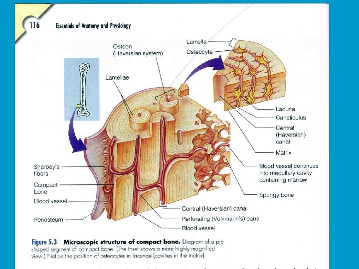
A structural unit of compact bone consisting central haversian canal. To know the structures of a synovial joint and a symphysis joint (intervertebral disc). Lesion composed of dense cortical bone (compact bone) with definite osteocyte lacunae and cement lines (line visible by microscopic examination marking the boundary of an osteon/ haversian system). The osteon consists of a central canal called the osteogenic (haversian) canal, which is surrounded by concentric rings (lamellae) of the matrix. Within these rings, are space called lacunae that contain osteocytes.
Bone Histology Types Of Bone Cells 1 Osteocyte from slidetodoc.com These cylinder shaped structures are called osteons or haversian systems. To know the architecture of compact and spongy (cancellous) bone. Osteoblasts deposit the matrix in the form of thin sheets which are called lamellae. Learn vocabulary, terms and more with flashcards, games and other study tools. Like compact bone, spongy bone, also known as cancellous bone, contains osteocytes housed in lacunae, but they are not arranged in concentric circles. The basic units of compact bone are called osteons or haversian systems. The osteon consists of a central canal called the osteogenic (haversian) canal, which is surrounded by concentric rings (lamellae) of the matrix. Between the rings of matrix, the bone cells (osteocytes) are located in spaces called lacunae.
The lacunae are situated between the lamellae, and consist of a number of oblong spaces.
The osteon consists of a central canal called the osteogenic (haversian) canal, which is surrounded by concentric rings (lamellae) of the matrix. In an ordinary microscopic section, viewed by transmitted light, they appear as fusiform opaque spots. The walls of the diaphysis are composed of dense and hard compact bone. The lacunae are situated between the lamellae, and consist of a number of oblong spaces. .cancellous vs compact bone diagram of a long bone withcompact and cancellous bone long bone with compact (arrows) and cancellous bone central canal and osteocytes in lacunae connected by canaliculi decalcified compact bone bones the cells of bone tem of an osteocyte in lacuane (left). Around the haversian canal, rings of bone tissue are found called lamellae. The musculoskeletal system is comprised of bones and connective tissue structures, such as cartilage, ligaments, and tendons. Compact bone consists of closely packed osteons or haversian systems. The osteon consists of a central canal called the osteonic (haversian) canal, which is surrounded by concentric rings (lamellae) of matrix. Compact bone consists of closely packed osteons or haversian systems. Compact bone is dense bone tissue found on the outside of a bone. Compact bone surrounds the spongy bone tissue and it has a unique appearance. A structural unit of compact bone consisting central haversian canal.
Lesion composed of dense cortical bone (compact bone) with definite osteocyte lacunae and cement lines (line visible by microscopic examination marking the boundary of an osteon/ haversian system) compact bone diagram. It can be remodeled all throughout life to withstand stress.